
Knee Yoga Anatomy & Function – How to protect during Yoga asana
The knee is the largest joint and one of the most important joints in the human body. It plays an essential role in the motion related to moving the body’s weight horizontally ( running and walking) and vertically ( jumping).

In humans and other primates, the knee connects the thigh to the leg and consists of two joints. Tibiofemoral joint ( which is femur and tibia joint) and other one is patellofemoral joint ( joint in between femur and patella).
It’s a hinge joint, type of synovial joint. which provides flexion and extension and slight internal and external rotation.
In between knee joint their is synovial fluid which is contained inside the synovial membrane called joint capsule.
Knee ligament:
Ligaments are bands of strong tissue that connect bone to bone. Knee has four ligaments which support knee for the move and help joint to be stable, connect femur and tibia together.
- Anterior cruciate ligament (ACL)
- Posterior cruciate ligament (PCL)
- Medial collateral ligament (MCL)
- Lateral collateral ligament ( LCL)
The ACL and PCL prevent the femur and tibia from sliding too far forward or backward. The MCL and LCL prevent side to side movement. A relatively new finding has revealed another ligament in the knee called the anterolateral ligament (ALL), which appears to function in conjunction with the ACL.
Ligaments and knee arthritis:
Injuries to the knee ligaments can lead to instability of the joints, which intensify the wear and tear that leads to arthritis of the knee. Conversely, knee arthritis can lead to joint instability, which puts more pressure on the ligaments and increases the risk of ligament injuries.
KNEE TENDON:
Tendon tissue connects bone to muscle. The largest tendon of the knee is the patellar tendon.
The patella tendon starts from the quadriceps muscle of the thigh and extends downward, attaching to the patella in front of the tibia. When the quadriceps muscles contract, the patellar tendon is pulled and the leg straightens.
Tendons and arthritis:
Patellar tendon problems can result from arthritis of the knee, but are more likely to affect athletes who do a lot of running, walking, and jumping. If the patellar tendon becomes irritated and inflamed it is called patellar tendinopathy, also known as jumper’s knee.
Knee Bursar:
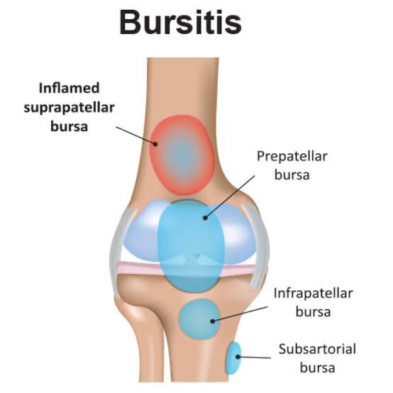
Bursa located between bone and soft tissues it’s a tiny, slippery, fluid- filled sac.
Like cartilage, the bursa reduces friction. But while cartilage reduces friction between bones, the bursa reduces friction between bones and soft tissues, such as muscles and tendons—the bursa helps muscles and tendons slide freely when the knee joint moves. There are many bursae in the knee.
The Knee’s Synovial Membrane and Synovial Fluid:
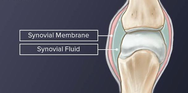
Synovial membrane surrounds the knee joint and produces synovial fluid that lubricates and nourishes the knee.
When the knee is at rest, synovial fluid is contained in the cartilage, just as water is in a sponge. When the knee is bent or bearing weight, synovial fluid leaks out. Hence the use of joints is essential to keep the joints lubricated and healthy.
Muscles of the Knee
Quadriceps: Represent the group of 4 muscles in the front thigh. These muscles are allowing the knee to straighten. This movement is essential for getting up from a sitting position, bringing your foot forward while walking, and kicking the ball!

Hamstrings: Is the group of 3 muscle which helps the knee to bend. These muscle helps to lifting the foot to walk.

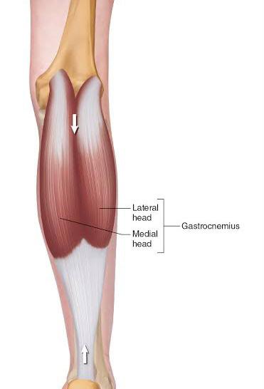
Gastrocnemius: It’s a group of 2 muscle which help the back lower leg (calf).
Get in Touch :
Yoga Teacher Training in Rishikesh 200 Hour Yoga Teacher Training in Rishikesh